Session 1 – Imaging
Wednesday, 10.9.2025, 2:30 pm – 3:45 pm
Chairs: Anja Hauser, Raluca Niesner
For a comprehensive understanding of molecular mechanisms of cellular function and dysfunction, in health and disease, information on the effects of microenvironmental cues in the tissue is needed. With the emergence of spatial biology technologies and analysis, we recently experienced a leap towards achieving this aim, whereas newest developments in optics and nanotechnology steadily improve our capacity to gain information about cells and tissues in vivo. In this session, Artür Manukian (Max Delbrück Center for Molecular Medicine, Berlin) will give us an overview on the development of algorithms to analyze multi-dimensional spatial omics data, also involving deep-learning-based analysis, and on numerical strategies to integrate the information from various spatial technologies. Luigi Bonacina (Applied Physics, University of Geneva) will focus in his talk on cutting-edge development of non-linear optical methods using nanocrystals for enhanced bioimaging in vivo. The session will be rounded up by short talks addressing strategies of DNA damage detection by Niklas Schmitt (OncoRay, Medical University, Dresden) and flow sorting of neutrophil granulocytes based on nuclear morphology by Pia Truebenbach (Fraunhofer Institute Potsdam).

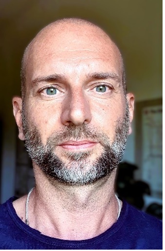
Luigi Bonacina
Nonlinear BioImaging LAB, Department de Physique Appliquée, Université de Genève
Geneva, Switzerland
Nanotechnology and Nonlinear Optics Strategies for Bioimaging
Multiphoton excited fluorescence is just one facet of the multiple signals that arise from the nonlinear optical interaction between light and matter. The acquisition of most of these nonlinear signals (harmonics, frequency mixings) in microscopy has been hindered so far by a combination of technological limitations, such as a lack of suitable optical components, and sample-related challenges, particularly the photostability of biological samples exposed to high peak power irradiation. The recent availability of rugged femtosecond optical parametric amplifiers (OPAs) operating in the Short-Wave InfraRed region (SWIR, 1-2 μm) at 1-10 MHz repetition rate, along with picosecond Fourier Domain Mode Locked (FDML) lasers have disclosed exciting new possibilities for bioimaging, offering improved contrast, penetration depth, and enabling fast volumetric imaging with minimal sample preparation requirements. I will present results from our work combining second and third harmonic generation from label-free samples and introduce an alternative diffractive-scanning approach to increase 3D acquisition speed. I will also illustrate how dielectric nanoparticles can be used to increase detection sensitivity on target structures facilitating high speed volumetric imaging. In conclusion, I will discuss the challenges associated with these imaging conditions in terms of sample photostability.
Biosketch
Luigi Bonacina (https://www.unige.ch/gap/nbi/ ) started his career at the University of Milan. After obtaining a Master in Optics, he moved to Switzerland where he completed a PhD at the Swiss Federal Institute of Technology of Lausanne (EPFL) with a research in the field of ultrafast molecular spectroscopy. Successively, he joined the Applied Physics Department of the University of Geneva where he is now Associate Professor. His research activities are focused on the development of novel bio-imaging approaches based on the combination of nanotechnology and nonlinear optics. Over time, Luigi Bonacina has established a dynamic and truly interdisciplinary network of European researchers as testified by several collaborative publications in the fields of cancer, regenerative, and circadian medicine which go along with articles in the fields of optics, spectroscopy, and nanotechnology. Since Jan 2022, he is coordinator of the EU H2020 project FAIR CHARM (www.faircharm.eu).

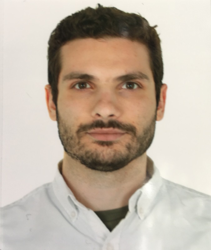
Artür Manukyan
Berlin Institute for Medical Systems Biology (BIMSB), Max Delbrück Center for Molecular Medicine, Berlin, Germany
VoltRon: A Spatial Omics Analysis Platform for Multi-Resolution and Multi-omics Integration using Image Registration
Rapid developments in spatial omics technologies have created a demand for computational platforms capable of analyzing spatial omics datasets with multiple modalities and diverse spatial resolutions. Meanwhile, bioimage analysis is reemerging as an integral part of spatial omics analysis where automated image registration and alignment approaches are essential for precise spatially aware data integration across tissue sections. To address this, we have developed VoltRon, a novel spatial omics integration platform that supports analysis of a large selection of spatial omics data modalities with comprehensive bioimaging data processing capabilities. To connect and integrate spatial multi-omics profiles across adjacent/serial tissue sections, VoltRon features scalable image registration workflows for automated synchronization of spatial coordinates and large images. This novel framework also accommodates omics data that are beyond single cell and spot level, thus allowing the analysis of molecules, regions of interests as well as image tiles/pixels that are often overlooked in spatial omics analysis frameworks. VoltRon integrates multiple modalities across tissue sections which we demonstrate by integrating the pathology driven analysis onto spatially localized SARS-CoV-2 viral RNAs within infected lung tissue, using image co-registration of histological images.
BioSketch
Artür Manukyan is a trained statistician, computational scientist and bioinformatician working at BIMSB, Berlin. He develops graph-based data integration algorithms and software platforms for spatial omic technologies, and he aims to combine these tools with foundation models capable of learning patterns from multiple omic and imaging modalities. He is also interested in biological knowledge graphs as well as other machine learning methods and software solutions to help overcoming computational challenges in translational medicine.
Niklas Schmitt
Schmitt N.1, Lange S.1,2, Wondrak M.1 & Kunz-Schughart L.A.1,3
1 OncoRay – National Center for Radiation Research in Oncology, Faculty of Medicine, Dresden, Germany, 2 DataMedAssist, HTW – University of Applied Sciences, Dresden, Germany, 3 National Center for Tumor Diseases (NCT), NCT/UCC Dresden, Germany
Pros & Cons: dSTRIDE vs. gH2AX for direct vs. indirect DNA damage detection
DNA is the main target in radiotherapy-induced cell death. Residual DNA double-strand breaks (DSBs) are crucial for the curative outcome and, therefore, used as an analytical endpoint in radiobiology. The current cell-based immunofluorescent approaches for quantifying DSBs are indirect assays that utilize biomarkers of the DNA damage response. One prime example is the DNA-associated phosphorylated histone H2AX, which allows the counting of irradiation-induced foci (IRIF). In 2020, a novel, direct technique for detecting DSBs was introduced, namely dSTRIDE (SensiTive Recognition of Individual DNA Ends for double DNA-strand breaks). Our study aimed to evaluate the use of dSTRIDE for directly assessing DNA damage upon irradiation via fluorescence microscopy and systematically compare it with a routine γH2AX assay. As the initial dSTRIDE procedure did not meet the requirements for IRIF detection, a modified dSTRIDE protocol will be presented. The adaptations enabled the visualization of DNA damage in two representative, differently radiosensitive human head and neck squamous cell carcinoma cell lines (FaDu, SAS) after increasing single-dose X-rays were delivered at a dose rate of 1.3 Gy/min using a YXLON Maxishot. By imaging and counting the foci of at least 500 cells per condition, we verified the clear dose-dependent radioresponses and assessed the time-kinetics of the DNA damage. Co-localization experiments for dSTRIDE and gH2AX foci are ongoing, as is the testing of the approach in tissue material beyond single cells. Based on our experiences, we will highlight the pros and cons of the approach.
Pia Truebenbach
Pia Truebenbacha, David Lallingera, Felix Pfisterera, Neus Godinoa and Michael Kirschbauma
aFraunhofer Institute for Cell Therapy and Immunology IZI, Branch Bioanalytics and Bioprocesses IZI-BB, Am Muehlenberg 13, 14476 Potsdam, Germany
Surface marker-free identification and flow sorting of neutrophil granulocytes based on nuclear morphology
Neutrophil granulocytes play a central role in innate immune defense but pose a particular challenge for experimental analyses due to their short lifespan and high sensitivity to external stimuli. Conventional isolation methods such as density gradient centrifugation, MACS or FACS often require large sample volumes or rely on staining with specific antibodies that lead to neutrophil activation via Fcy receptors and ultimately cell death1. However, neutrophils have a specific, lobulated nuclear morphology that allows them to be identified in the microscope image without markers via the nuclear circularity value. Since they tolerate staining with nucleic acid-binding dyes such as DRAQ5, we used this for marker-free and thus stress-reduced sorting with a microfluidic image-activated cell sorting method.2, 3
To this end, staining protocols for the cells and their nuclei were first developed in order to create a (fluorescence) image database that served as the basis for finding the classification algorithm. Using a confusion matrix, several circularity thresholds were evaluated in terms of their discrimination efficiency. The algorithm was included as a classifier in our image-based cell sorter and used for isolating neutrophils from erythrocyte-depleted capillary blood samples. In particular, with a circularity threshold value of 0.70, a neutrophil purity of up to 91 % was achieved with a recovery efficiency showing no evidence of significant systematic cell damage or cell losses. The results presented in this paper thus contribute to the development of gentle, marker-free cell sorting systems and open up new perspectives in the functional analysis of neutrophil granulocytes.
References:
1 A. N. Connelly et al., Sci Rep, 2022, 12(1):3667.
1 T. Gerling et al., Lab Chip, 2023, 23, 3123-3302
2 N. Godino et al., Lab Chip, 2019, 19, 4016–4020.
