Klaus Goerttler Session
Wednesday, 10.09.2025, 4:15 pm – 6:00 pm
Chair: Leonie Kunz Schughart, Dresden

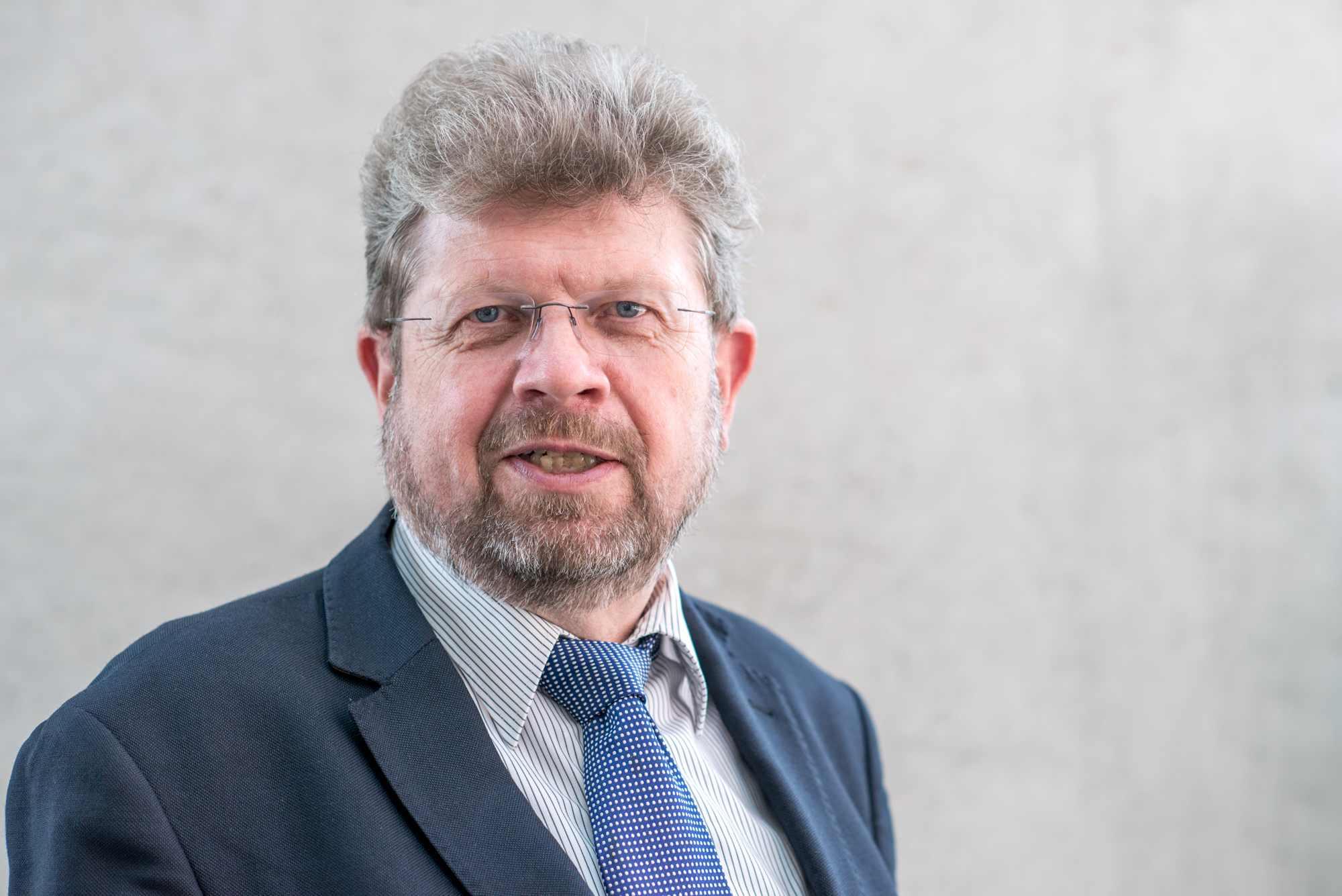
Prof. Dr. rer. nat. Andreas Radbruch
German Rheumatology Research Center
a Leibniz Institute, Berlin
Cells in the Limelight
Robert Hooke coined the term “cell” in 1665, but it took until the mid-19th century to realize that cells are the basic compartments of life and disease. Microscopy was the tool for discovery and sketches for documentation. It took another 100 years until electron-microscopes detailed out the internal structures of cells. Soon after, in 1953, the impedance-based flow cytometer of Wallace Coulter allowed to count and size large numbers of individual cells, and in 1968, Wolfgang Göhde from Münster developed the first fluorescence-based flow cytometer. It was designed to detect (rare) tumor cells with aberrant DNA content. Karyotyping by cytometry was the major driving force of the technology at that time, with prominent labs in Los Alamos and Lawrence Livermore in the US. Cytometry took another twist, when fluorescent dyes were combined with monoclonal antibodies, first described in 1975, to label cells for distinct expression of proteins. At about the same time, Mack Fulwyler and Len Herzenberg developed the first fluorescence-activated cell sorter (FACS), allowing to isolate cells with a distinct phenotype. I joined the field in 1976, lucky in retrospect, as a student in Klaus Rajewsky´s lab at the Cologne University. We had a FACS, made monoclonals and analysed how B cells made antibodies. The combination of immunofluorescence, monoclonals and cytometry shifted the emphasis of cytometry from one-color karyotyping to multiparametric proteomics, driving also the development of fluorescent biochemical applications, with the Valet group in Munich as an international lead. It should be noted that cytometers of those days required some expertise in operation, many of todays “easy-to-use” options had not been developed, clogging and realignment remained a constant companion during operation, personal computers entered the scene only about 10 years later. The vendors did not have an established service. The need for communication was obvious, on the national and international level. The cytometric centers of Germany started to organize events for exchange of expertise, in Munich, Cologne, Regensburg, Münster, Heidelberg etc. Although working in different areas of engineering, operation, and biomedicine, there was a mutual agreement that it would be good to have an annual meeting, easy to access: In 1990 the DGfZ was founded. Klaus Goerttler was able to secure the DKFZ meeting venue in Heidelberg for the initial years, and from then on, the DGfZ continued to be the place where you meet to discuss cytometry. It is interesting that till today the weakest point of cytometry is the human user, caring for the cells and interpreting the data. From my perspective, the development of flow cytometry has reached a certain ceiling with respect to mass and spectral cytometry maximizing the number of parameters per cell, access to intracellular and secreted proteins and to many other parameters, large data storage and evaluation capacities, streamlined operation, competent service. FACS and MACS are still the technologies of choice for the isolation of cells-of-interest. While in the founding years cells, mostly from blood, were considered as more or less independent units of life, today we realize that they are controlled by their environment. The inclusion of sophisticated microscope technologies for spatial cytometry is mandatory and a most valuable asset of the DGfZ. There also would be reasons to include single cell transcriptomics, methylomics, metabolomics, microbiomics etc. into our spectrum of technology, to transform the DGfZ into the forum for looking at cells.
Biosketch
A biologist by education, Andreas Radbruch has focussed his scientific curiosity on the immune system, and in particular immunological memory, and the way it provides immunity and generates immunopathology.
Andreas Radbruch obtained his PhD at the Genetics Institute of the Cologne University, Germany, with Klaus Rajewsky in 1980. He later became Associate Professor there and was a visiting scientist with Max Cooper and John Kearney at the University of Alabama, Birmingham. In 1996, he became Director of the German Rheumatology Research Center in Berlin, now a Leibniz Institute (until 2023), and in 1998, Professor of Rheumatology at the Charité Medical Center and Humboldt University of Berlin.
Andreas Radbruch has developed a line of research aiming at a cellular and molecular understanding of immune reactions and immunological memory. His research approach is based on the analysis of individual cells, developing and using cutting-edge technologies for cytometry and cell sorting. His lab developed the MACS technology, cytokine cytometry, the cytometric secretion assay, magnetofluorescent liposomes and other tools to analyze fate decisions and imprinting of lymphocytes. He initially focused on the transcriptional regulation of antibody class switch recombination, the shaping of antibody specificity by somatic mutation in B lymphocytes, and the epigenetic imprinting of cytokine gene expression in T lymphocytes. The discovery of long-lived (memory) plasma cells in 1997 initiated a line of research that so far has generated a new understanding of immunological memory, as maintained by functionally imprinted memory plasma cells, and memory B and T lymphocytes, which, as this group has shown, individually reside, rest and survive in niches provided by mesenchymal stromal cells, mostly in the bone marrow, but also in other tissues. Of clinical relevance, memory plasma cells secreting pathogenic antibodies have been recognized as being refractory to conventional therapies and a critical and so far unmet therapeutic target in antibody-mediated diseases. The same is true for memory T lymphocytes driving chronic inflammation, for which the group has identified molecular adaptations and novel therapeutic targets, like the transcription factor Twist1, which dampens pathogenicity and promotes persistence of Th lymphocytes in chronic inflammation. Andreas Radbruch has received various scientific awards and honors, the Carol Nachman Prize for Rheumatology (2011), an Advanced Grant of the European Research Council (ERC, 2011), and the Avery Landsteiner Award (2014). He is a member of the Berlin Brandenburg Academy of Sciences, the European Molecular Biology Organization (EMBO) and the Leopoldina, the German National Academy of Sciences. He has been president of the International Society for the Advancement of Cytometry (ISAC; 2014-2016), of the German Societies for Immunology (2009-2010) and for Rheumatology (2007-2008), and of the European Federation of Immunological Societies (EFIS) (2019 -2021).

Lisa Budzinski
Berlin
Abstract
Microbiome research is a promising field in human medicine, exploring the microbial communities that inhabit the human body and their potential to influence health. Advances in sequencing technologies and bioinformatics have greatly expanded our understanding of the microbiome’s composition and potential functions.
To investigate microbiome–host interactions more directly, we developed a single-cell analysis approach: microbiota phenotyping. This tool profiles microbial communities for their cellular properties, examines their interactions with the human immune system, and analyzes the resulting complex datasets using machine learning.
I will present examples from chronic inflammatory diseases, mainly inflammatory bowel and rheumatic diseases, where we observed patterns of altered microbial phenotypes. These patterns reveal the dynamic nature of human microbial communities and their cellular-level interactions with the host, and, when combined with machine learning, show strong potential for diagnostics, patient stratification, and therapy monitoring.
In summary, microbiota phenotyping adds a valuable single-cell perspective to microbiome research and opens new opportunities for clinical applications.
Short-Bio
Lisa Budzinski studied Biotechnology at TU Berlin and has focused on single-cell analysis since her Master’s project on mass cytometry, conducted at the German Rheumatology Research Centre Berlin (DRFZ), where she developed a quality assurance protocol.
Shifting from mass to flow cytometry and from human to bacterial cells, she dedicated her PhD thesis to developing a multiparametric single-cell analysis tool for the human intestinal microbiota in the Schwiete Laboratory for Microbiota and Inflammation at DRFZ Berlin. During her PhD, she actively engaged with the national and international cytometry community, attending numerous conferences and meetings, and receiving several awards for her work. Alongside her passion for science and innovation, Lisa is committed to science communication in various formats, advocating for science in society and making knowledge accessible

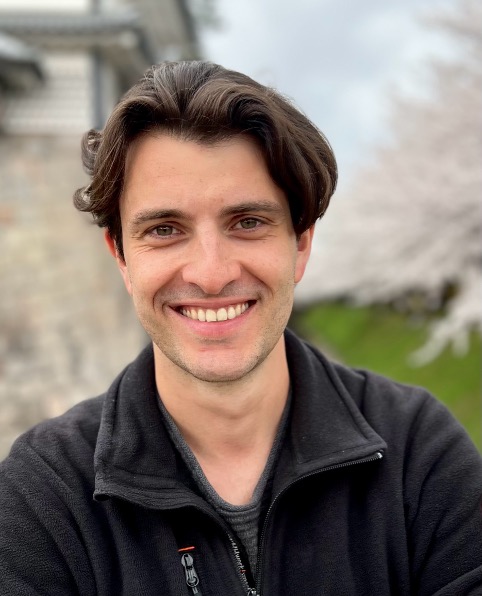
Tobias Kammann
Basel
Charting the diversity of MAIT cells across the human body
Mucosal-associated invariant T (MAIT) cells are unconventional lymphocytes that recognize microbial riboflavin metabolites via MR1, yet their roles in human tissue immunity remain unclear. We profiled MAIT cells in blood, barrier and lymphoid organs from healthy human donors, revealing frequencies that vary markedly by donor and site. Immunophenotyping further revealed site-specific adaptation of MAIT cells: In the intestines, CD103⁺ MAIT cells adopt an immunoregulatory CD39+ profile. In the liver, CD56⁺ MAIT cells exhibit enhanced effector capacity, innate-like behaviour, and polyfunctionality. Both signatures accumulated with age, suggesting tissue microenvironmental “training”. Moreover, the CD56+ profile was inducible on CD56⁻ MAIT cells by exposure to antigen or IL-7 in vitro, highlighting functional plasticity of MAIT cells. In summary, we demonstrate functional heterogeneity and site-specific adaptation in resident MAIT cells across human barrier tissues with distinct regulatory and effector signatures.
Biosketch
Tobias Kammann is an immunologist (PhD), biochemist (BSc and MSc), and self-trained computational biologist. His research explores how human T cell immunity is tuned by the tissue microenvironment and by training. After undergraduate education in biochemistry with a focus on immune-metabolic sepsis research at the Friedrich Schiller University in Jena, Germany, Tobias completed his PhD in T cell immunology at the Center for Infectious Medicine at the Karolinska Institute in Stockholm in December 2024. In his PhD, Tobias first contributed to Sweden’s COVID-19 research response and later focussed on exploring the diversity of mucosal-associated invariant T cells across human tissues. After a brief postdoc at the Karolinska Institute where he extended his exploration to other tissue-resident T cell populations, Tobias joined Roche in Basel, Switzerland, as a postdoctoral fellow in Immunology Discovery.
