
Klaus Goerttler Keynote
Ulrich F. Keyser - Nanopore microscopy: Towards single-cell readout of RNA with single-molecule sensitivity
Chair: Oliver Otto
Speaker: Ulrich F. Keyser
Cavendish Laboratory, University of Cambridge, UK
Biosensing with nanopores is developing rapidly with commercial DNA and RNA sequencing platforms challenging established next generation sequencing technologies. Nanopores offer the unique ability to translate molecular structure directly into electrical signals without the need of fluorescent labels. Beyond sequencing of nucleic acids and proteins, many other applications are enabled by counting single molecules with nanopores.
First, I will describe the identification of long transcript isoforms at the single-molecule level using solid-state nanopore microscopy. We refold target RNA into RNA identifiers (IDs) with designed sets of complementary DNA strands. Each reshaped molecule carries a unique sequence of structural (pseudo)colours. The sequence of structural colours of RNA identifiers enables simultaneous identification and relative quantification of multiple RNA targets without prior amplification. RNA IDs discriminate circular and linear transcript isoforms in a one-step, enzyme-free reaction in a complex human transcriptome with single-molecule resolution [1]. We will show recent results on detection of RNA modifications like 5mC, Inosine and MeC using RNA nanotechnology in ribosomal RNA (rRNA) from pathogenic bacteria like A. baumannii [2]. Finally, I will discuss how the technique may enable single-cell RNA detection by adapting the process for integration into droplet microfluidics.
References:
- Bošković and U. F. Keyser. Nanopore microscope identifies RNA isoforms with structural colors. Nature Chemistry, 14:1258-1264, 2022.
- Li, S. C. Meng, Y. Wang, C. M. Platnich, M. K. Earle, E. Mylona, P. Naydenova, S. Baker, J. Zhu and U. F. Keyser. Nanopore detection of single-nucleotide RNA mutations and modifications with programmable nanolatches. Nature Nanotechnology, published online, 2025.
Ulrich F. Keyser
Cavendish Laboratory, University of Cambridge, UK
Nanopore microscopy:
Towards single-cell readout of RNA with single-molecule sensitivity
Biosensing with nanopores is developing rapidly with commercial DNA and RNA sequencing platforms challenging established next generation sequencing technologies. Nanopores offer the unique ability to translate molecular structure directly into electrical signals without the need of fluorescent labels. Beyond sequencing of nucleic acids and proteins, many other applications are enabled by counting single molecules with nanopores.
First, I will describe the identification of long transcript isoforms at the single-molecule level using solid-state nanopore microscopy. We refold target RNA into RNA identifiers (IDs) with designed sets of complementary DNA strands. Each reshaped molecule carries a unique sequence of structural (pseudo)colours. The sequence of structural colours of RNA identifiers enables simultaneous identification and relative quantification of multiple RNA targets without prior amplification. RNA IDs discriminate circular and linear transcript isoforms in a one-step, enzyme-free reaction in a complex human transcriptome with single-molecule resolution [1]. We will show recent results on detection of RNA modifications like 5mC, Inosine and MeC using RNA nanotechnology in ribosomal RNA (rRNA) from pathogenic bacteria like A. baumannii [2]. Finally, I will discuss how the technique may enable single-cell RNA detection by adapting the process for integration into droplet microfluidics.
References:
- Bošković and U. F. Keyser. Nanopore microscope identifies RNA isoforms with structural colors. Nature Chemistry, 14:1258-1264, 2022.
- Li, S. C. Meng, Y. Wang, C. M. Platnich, M. K. Earle, E. Mylona, P. Naydenova, S. Baker, J. Zhu and U. F. Keyser. Nanopore detection of single-nucleotide RNA mutations and modifications with programmable nanolatches. Nature Nanotechnology, published online, 2025.
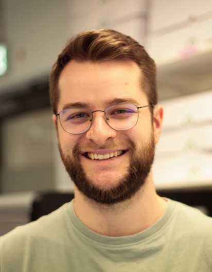
Tobias Walther
DNA hydrogel microparticles as phantom cells for tissue engineering
Short Abstract
What if we could mimic cellular functions with synthetic materials? Hydrogel microparticles (HMPs) allow for exactly that by forming cell-sized particles with controllable mechanics and the ability for chemical signalling through stimuli-response. This way, HMPs become functional cell mimics: phantom cells. As a material, DNA is especially promising for the creation of phantom cells due to its simple sequence design, viscoelastic mechanics and complex stimuli response. Here, we showcase that DNA sequence design can be used to create cell-sized DNA-HMPs with finely programmed viscoelasticity, mimicking cellular mechanics. Using click chemistry, the DNA-HMPs are modified to physically interact with living cells and act as force sensors, while light-triggered morphogen release allows to direct stem cell differentiation in organoids. DNA-HMPs thus present a versatile tool to probe and manipulate tissue behaviour in multicellular systems.
Abstract
Hydrogel microparticles (HMPs) are powerful tools for the study and manipulation of 3D cellular systems. Engineered to allow physical interactions with living cells and chemical signalling through external triggers, they can be directly interfaced with growing tissues and act as functional cell mimics: phantom cells. Most polymeric HMPs, however, lack versatility in terms of their capacity for targeted chemical functionalization combined with fine-scale mechanical programmability, limiting their complexity. DNA-based materials offer promising properties in this regard due to their sequence-defined self-assembly, precise mechanical tuning, and straightforward chemical modification. Here, we introduce fully DNA-based hydrogel microparticles (DNA-HMPs) as a new class of multifunctional phantom cells for tissue engineering1,2,3. DNA-HMP size can be controlled via microfluidics to span the size range of human tissue cells. Making use of DNA sequence design, DNA-HMPs demonstrate finely programmable mechanics with viscoelastic behaviour and Young’s Moduli between 30 Pa – 6 kPa, as validated by real-time deformability cytometry and indentation. This way, their mechanical properties can be tailored to mimic specific cell types of target tissues. DNA-HMP modification based on click chemistry further enables controlled physical interactions with living cells for force sensing in 3D1, as well as binding of morphogenic compounds. Through light-based stimuli-response, these morphogens can be released from the DNA-HMPs to spatiotemporally control stem cell differentiation in organoids, increasing cell type diversity2. Combining programmable material properties with straightforward functionalization and stimuli-responsiveness, DNA-HMPs represent a versatile new tool to probe and manipulate tissue behaviour in 3D cell culture, advancing tissue engineering through DNA nanotechnology.
1: Walther T et al. (2026) Advanced Materials 38, no. 37: e14218, doi.org/10.1002/adma.202514218
2: Afting C*, Walther T* et al. (2024) Nature Nanotechnology 19, 1849–1857, doi.org/10.1038/s41565-024-01779-y
3: Brauburger S*, Kraus BK*, Walther T* et al. (2026) Soft Matter, 22 (30): 4978–4993, doi.org/10.1039/d6sm00242k
*: Authors contributed equally
Biosketch
Tobias Walther is a molecular biotechnologist (B.Sc./M.Sc.) with a PhD in Molecular Systems Science and Engineering from Heidelberg University. In the lab of Prof. Dr. Kerstin Göpfrich at the Max Planck Institute for Medical Research and the Center for Molecular Biology of Heidelberg University (ZMBH), his PhD focused on the engineering of biomimetic materials and synthetic cellular systems. He used DNA as a programmable material to create DNA-based hydrogel microparticles as phantom cells – biophysical mimics of living cells capable of controlled chemical signalling – directing and understanding cellular behaviour in engineered tissues. With his research, Tobias merges DNA nanotechnology, cell biology, cytometry, biomaterials design and bio-orthogonal chemistry to develop new methods to study and engineer complex cellular environments. Currently, he is a postdoctoral researcher in the group of Prof. Dr. Manuel Salmeron-Sanchez at the Institute for Bioengineering of Catalonia (IBEC) where he works on new biomaterials for disease modelling.